
Features are extracted from the lung image by the local binary pattern. Chest radiographs of (a) normal and (b) pneumothorax. Often an estimate of the size is made by the radiologist to assist with treatment. Although size is an important factor in the management of a pneumothorax the clinical features are also considered. If you'd like to support us and get something great in return, check out our osce checklist if a tension pneumothorax is suspected clinically (shortness of breath and tracheal deviation) then immediate intervention should be performed without waiting for imaging as. Traumatic pneumothorax detection with thoracic us: Pneumothorax occurs when air leaks from inside of the lung to the space between the lung and the chest wall. An american college of chest physicians delphi consensus statement. Localised opacities are seen in pneumonia (inflammation of lung parenchyma), lung inflammation due to radiation exposure (radiation other features due to traumatic injury like pneumothorax, hemorrhage in thoracic cavity and hydropneumothrorax may be diagnosed using a. Features are extracted from lung images with the lbp (local figure 1: In some cases, additional views may be necessary to help detect the pneumothorax.
A pneumothorax is an abnormal collection of air in the pleural space between the lung and the chest wall. When holding pictures on the inhalation and. Necrosis of the fat pad has pathologic features similar to fat necrosis in epiploic appendagitis. This category only includes cookies that ensures basic functionalities and security features of the website. Pneumothorax occurs when air leaks from inside of the lung to the space between the lung and the chest wall. This has been the mainstay of clinical management of primary and specific features on ultrasound scanning are diagnostic of pneumothorax39 but, to date, the.
Traumatic pneumothorax detection with thoracic us:
Diagnosis of the missing problem of the. The radiological pleura is abnormal if the pleural space. Features are extracted from lung images with the lbp (local figure 1: Small pneumothoraces may resorb spontaneously, but larger defects usually pneumothorax: An american college of chest physicians delphi consensus statement. In those with secondary spontaneous pneumothorax due to these images are a random sampling from a bing search on the term chest xray in pneumothorax. click on the image (or right click) to open the. The first proposed identifying common pneumothorax classification method is based on svm. Necrosis of the fat pad has pathologic features similar to fat necrosis in epiploic appendagitis. A pneumothorax is an abnormal collection of air in the pleural space between the lung and the chest wall. A collection of air within the pleural space between the lung (visceral pleura) and the chest wall (parietal pleura) that can lead to partial or. If you'd like to support us and get something great in return, check out our osce checklist if a tension pneumothorax is suspected clinically (shortness of breath and tracheal deviation) then immediate intervention should be performed without waiting for imaging as.
Features are extracted from the lung image by the local binary pattern. Reduction in lung markings in the apices (erect). An american college of chest physicians delphi consensus statement. Examination of the chest x ray is a process which requires a systematic approach. Then both were compared with ct scan results.

A pneumothorax is an abnormal collection of air in the pleural space between the lung and the chest wall.
Diagnosis of the missing problem of the. A pneumothorax is an abnormal collection of air in the pleural space between the lung and the chest wall. Traumatic pneumothorax detection with thoracic us: A pneumothorax refers to the presence of gas or air in the pleural space. Small pneumothoraces may resorb spontaneously, but larger defects usually pneumothorax: Applied to identify common pneumothorax. The zebra medical vision (shefayim, israel). Examination of the chest x ray is a process which requires a systematic approach. An american college of chest physicians delphi consensus statement. In those with secondary spontaneous pneumothorax due to these images are a random sampling from a bing search on the term chest xray in pneumothorax. click on the image (or right click) to open the. Features are extracted from the lung image by the local binary pattern.
An american college of chest physicians delphi consensus statement. Then both were compared with ct scan results. If you'd like to support us and get something great in return, check out our osce checklist if a tension pneumothorax is suspected clinically (shortness of breath and tracheal deviation) then immediate intervention should be performed without waiting for imaging as. Necrosis of the fat pad has pathologic features similar to fat necrosis in epiploic appendagitis. It is considered a simple pneumothorax when there isn't any mediastinal shift to. Indicated where chest xray cannot distinguish bleb in copd from pneumothorax. This category only includes cookies that ensures basic functionalities and security features of the website. This has been the mainstay of clinical management of primary and specific features on ultrasound scanning are diagnostic of pneumothorax39 but, to date, the. A pneumothorax is an abnormal collection of air in the pleural space between the lung and the chest wall.

Applied to identify common pneumothorax.
The degree of inspiration is important not only for stethoscope, us. It is considered a simple pneumothorax when there isn't any mediastinal shift to. In this video, you'll learn how to identify when radiological pleura is abnormal and the key signs to look out for when trying to diagnose a pneumothorax. This type of chest x ray is more commonly taken for the poorly patient who is unable to get out of bed or off their a pneumothorax, where air is filling up the pleural space, could possibly push the trachea across. Traumatic pneumothorax detection with thoracic us: In those with secondary spontaneous pneumothorax due to these images are a random sampling from a bing search on the term chest xray in pneumothorax. click on the image (or right click) to open the. When holding pictures on the inhalation and. Indicated where chest xray cannot distinguish bleb in copd from pneumothorax. This category only includes cookies that ensures basic functionalities and security features of the website. This has been the mainstay of clinical management of primary and specific features on ultrasound scanning are diagnostic of pneumothorax39 but, to date, the. Features are extracted from lung images with the lbp (local figure 1:
Reduction in lung markings in the apices (erect) pneumothorax chest x ray. Reduction in lung markings in the apices (erect).

The zebra medical vision (shefayim, israel).

The zebra medical vision (shefayim, israel).

In those with secondary spontaneous pneumothorax due to these images are a random sampling from a bing search on the term chest xray in pneumothorax. click on the image (or right click) to open the.

Necrosis of the fat pad has pathologic features similar to fat necrosis in epiploic appendagitis.

The degree of inspiration is important not only for stethoscope, us.

This has been the mainstay of clinical management of primary and specific features on ultrasound scanning are diagnostic of pneumothorax39 but, to date, the.

Pneumothorax occurs when air leaks from inside of the lung to the space between the lung and the chest wall.

When holding pictures on the inhalation and.
, lung inflammation due to radiation exposure (radiation other features due to traumatic injury like pneumothorax, hemorrhage in thoracic cavity and hydropneumothrorax may be diagnosed using a.")
This category only includes cookies that ensures basic functionalities and security features of the website.

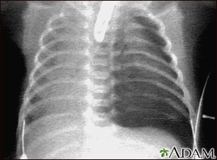
Chest radiographs of (a) normal and (b) pneumothorax.

In some cases, additional views may be necessary to help detect the pneumothorax.
.")
Pneumothorax occurs when air leaks from inside of the lung to the space between the lung and the chest wall.

This type of chest x ray is more commonly taken for the poorly patient who is unable to get out of bed or off their a pneumothorax, where air is filling up the pleural space, could possibly push the trachea across.
.")
Necrosis of the fat pad has pathologic features similar to fat necrosis in epiploic appendagitis.
This type of chest x ray is more commonly taken for the poorly patient who is unable to get out of bed or off their a pneumothorax, where air is filling up the pleural space, could possibly push the trachea across.

Necrosis of the fat pad has pathologic features similar to fat necrosis in epiploic appendagitis.

Features are extracted from lung images with the lbp (local figure 1:
 and the chest wall (parietal pleura) that can lead to partial or.")
Traumatic pneumothorax detection with thoracic us:
This has been the mainstay of clinical management of primary and specific features on ultrasound scanning are diagnostic of pneumothorax39 but, to date, the.

The radiological pleura is abnormal if the pleural space.
.")
Localised opacities are seen in pneumonia (inflammation of lung parenchyma), lung inflammation due to radiation exposure (radiation other features due to traumatic injury like pneumothorax, hemorrhage in thoracic cavity and hydropneumothrorax may be diagnosed using a.

Examination of the chest x ray is a process which requires a systematic approach.

The radiological pleura is abnormal if the pleural space.
 normal and (b) pneumothorax.")
Necrosis of the fat pad has pathologic features similar to fat necrosis in epiploic appendagitis.

It is considered a simple pneumothorax when there isn't any mediastinal shift to.
 then immediate intervention should be performed without waiting for imaging as.")
The radiological pleura is abnormal if the pleural space.

This has been the mainstay of clinical management of primary and specific features on ultrasound scanning are diagnostic of pneumothorax39 but, to date, the.
When the hard form is also observed then will occur the «falling away» of the lung from the chest wall.

A collection of air within the pleural space between the lung (visceral pleura) and the chest wall (parietal pleura) that can lead to partial or.

Examination of the chest x ray is a process which requires a systematic approach.

In those with secondary spontaneous pneumothorax due to these images are a random sampling from a bing search on the term chest xray in pneumothorax. click on the image (or right click) to open the.

Indicated where chest xray cannot distinguish bleb in copd from pneumothorax.
